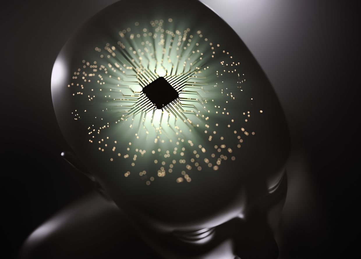
Scientists led by Xu Xiaomin from China have achieved a significant breakthrough in brain implant technology, creating an electrode array so thin and flexible that it matches the softness of brain tissue itself. The innovation, published in the peer-reviewed journal PNAS on April 28, addresses a problem that has constrained the development of reliable brain-computer interfaces for decades: the fundamental incompatibility between rigid electrodes and delicate neural tissue. Animal trials demonstrated that the new implant can record neural activity with exceptional clarity while remaining safely functional inside the body for at least 18 months, a duration that far exceeds conventional systems.
The challenge of building long-lasting brain implants has always centred on a seemingly insurmountable physics problem. Invasive electrode arrays, typically fabricated from platinum or platinum-iridium alloys, deliver the clearest neural signals available to researchers because they sit directly on the brain's surface. Yet their rigidity creates an inherent mismatch with the soft tissue environment they inhabit. Over months and years of implantation, this mismatch generates friction and microscopic movement at the electrode-tissue interface, triggering chronic inflammatory responses. The body's natural healing process then wraps scar tissue around the implants, progressively degrading signal quality until the system becomes unreliable.
The Chinese team's solution involves a material called conductive hydrogel with interfacial percolation, or Chip, which combines the electrical properties necessary for neural recording with the mechanical softness of brain tissue itself. This represents a fundamental departure from traditional electrode design. The hydrogel achieves exceptional electrical conductivity of up to 2,512 S/cm, enabling the transmission of faint neural signals with high fidelity. Yet conductivity alone does not solve the biocompatibility problem. The researchers faced another obstacle: conventional hydrogels absorb bodily fluids and expand, warping the precise microelectrode patterns built into them and ruining the carefully engineered spacing between channels.
To overcome this expansion problem, the team developed an innovative manufacturing approach using a rigid parylene substrate as a constraint frame. The hydrogel is pre-anchored to this substrate before processing, which prevents lateral expansion during fabrication. Working in the dry state, researchers then applied high-precision photolithography to create the electrode patterns. This method ensured that the hydrogel's structure remained stable throughout manufacturing, preserving the integrity of the electrode array. The final product is a 128-channel electrocorticography array measuring just nine micrometres in thickness—thinner than a human hair—with an exceptional channel density of 853 channels per square centimetre. This represents more than a tenfold improvement over previous hydrogel-based designs, allowing researchers to record neural activity from a much larger population of brain cells simultaneously.
The implications of this density increase are substantial for understanding brain function. Higher channel counts mean finer spatial resolution, enabling scientists to map neural activity across brain regions with greater precision. For clinical applications, this translates to more detailed information about the location and nature of neurological disorders, potentially improving diagnostic accuracy and treatment targeting. The electrode array's mechanical properties proved equally impressive in laboratory testing. When subjected to repeated stretching of up to 30 per cent—representing the maximum deformation that brain tissue typically experiences—the material maintained stable electrical performance with less than 4 per cent variation after 1,000 cycles. This durability suggests the implants can withstand the natural movement and deformation of living brain tissue without degradation.
Biocompatibility testing revealed another critical advantage. When researchers placed the electrode array directly on fresh porcine brain tissue in vitro, it conformed gently to the curved surface of the brain rather than sitting rigidly atop it. Crucially, the array could be peeled away without causing any tissue damage, indicating excellent interfacial adhesion and minimal mechanical trauma. These properties matter enormously for surgical implantation, as they suggest the procedure would be gentler and less likely to create micro-injuries that trigger inflammation. The hydrogel's ability to move with the brain rather than against it mirrors the behaviour of tissue naturally compatible with the neural environment.
Long-term animal studies provided the most compelling evidence of the technology's potential. The research team implanted Chip-based electrode arrays into five rabbits and recorded continuously for more than 550 days as the animals moved freely in their enclosures. Throughout this extended period, the signal-to-noise ratio—a key measure of recording quality—remained at least 94 per cent of its initial value. This consistency represents a dramatic improvement over conventional rigid electrodes, which typically show declining signal quality within months. Histological examination of brain tissue samples collected 16 weeks after implantation revealed minimal inflammatory response, confirming that the system does not provoke the chronic immune reaction that degrades conventional implants.
The significance of this work extends beyond the laboratory to patients with severe neurological conditions and paralysis. Brain-computer interfaces capable of reliable long-term operation could eventually enable paralysed individuals to control robotic limbs or computer cursors with their thoughts, restoring a degree of independence and agency. Current technology falls short because signal degradation forces frequent surgical replacement, an invasive and expensive procedure. A truly long-lasting implant that maintains signal quality for years would transform clinical possibilities. The same technology could also enhance deep brain stimulation therapies for Parkinson's disease, epilepsy, and depression, allowing more precise targeting and adjustment of treatment parameters.
For the broader field of bioelectronics, the researchers emphasise that their methods could extend far beyond neural interfaces. The team suggests that the manufacturing techniques and material innovations developed for brain implants could improve other bioelectronic systems that operate within the body's fluid environment. Sensors for monitoring heart function, implantable drug delivery systems, and devices for interfacing with peripheral nerves might all benefit from similar approaches. The hydrogel platform represents a new paradigm for creating soft, flexible bioelectronics that integrate harmoniously with living tissue rather than imposing mechanical incompatibility.
For Malaysia and Southeast Asia, this development carries particular relevance. The region faces a rising burden of neurological diseases including stroke, traumatic spinal cord injury, and neurodegenerative disorders, while access to advanced medical technology remains uneven. If this technology matures and becomes clinically available, it could eventually provide patients in the region with treatment options previously limited to wealthy countries. The research also highlights China's growing leadership in biomedical engineering and materials science, challenging the historical dominance of Western research institutions in medical device innovation. Malaysian scientists and institutions increasingly collaborate with Chinese counterparts, and developments like this open new possibilities for regional research partnerships and technology transfer. The work demonstrates how advances in fundamental materials science can translate into concrete health benefits, suggesting pathways for Southeast Asian nations to develop their own bioelectronics capabilities.